
Dog health: orthopaedic
Legg-Calve-Perthes disease in dogs: the toy-breed limp, the surgery that fixes it, and what it costs in India
Ten breeds with the number of dogs actually evaluated in the OFA register, the age window seven bodies draw differently, the bilateral proportion only one source prints, four outcome series with their denominators, and the Indian rate cards that price the surgery at a tenth of a private quote.
Quick answer
Legg-Calve-Perthes disease in dogs is the death of the ball of the hip in a small-breed puppy aged four to twelve months, after its blood supply fails. One hip is affected in most dogs and both in 12 to 16.5 percent. Treatment is surgical, removing the femoral head and neck so a false joint forms. An Indian government veterinary college prices a major operation at Rs 2,500 and a private chain prices this surgery at Rs 25,000 to Rs 60,000. Your nearby vet decides.
This guide is general information for Indian pet owners and is not a diagnosis. Always consult a registered veterinarian near you before acting on anything you read here, and treat any sudden change in your pet's condition as urgent.
Signs of Legg-Calve-Perthes disease in dogs
The 8 signs to watch for are listed below. Any one of them is a reason to consult your nearby vet; none of them is a diagnosis.
- A limp on one hind leg that gets worse over weeks, not better
- Reluctance to jump onto furniture or to climb stairs
- Thigh muscle visibly thinner on the affected side
- Pain or yelping when the hip is extended or rotated
- A grinding or crunchy feel in the hip during movement
- New irritability when the back end is touched or handled
- Carrying the leg, or refusing to put the foot down at all
- The affected leg looking shorter than the other one
Breeds on this site that document this condition
Each breed page below carries the figure for this condition with its denominator and its source. The 10 breeds listed are the ones on this site that document it, not a ranking of risk.
India has no published veterinary record of Legg-Calve-Perthes disease in a dog. Two Europe PMC instruments run on 27 September 2026 both returned zero, a title and abstract search and an author-affiliation search, while their canine controls returned 411 and 1,523 records, so the instrument was working. The same affiliation search without the dog clause returns 76 Indian papers on the disease in children. Meanwhile the breeds it targets, the Pomeranian, Pug, Lhasa Apso, Shih Tzu and terrier types, are exactly what an Indian city flat holds. A diagnostic radiograph costs Rs 150 at ICAR-IVRI, Rs 300 at Madras Veterinary College and Rs 400 at Bihar Animal Sciences University, against Rs 500 to Rs 5,000 privately, and the surgery runs Rs 2,500 to Rs 3,000 as a major operation at a government college against Rs 25,000 to Rs 60,000 at a private chain.
What Legg-Calve-Perthes disease in dogs is, and which dogs get it
Legg-Calve-Perthes disease in dogs is the death of the ball of the hip joint in a young small-breed dog, and it happens because the blood supply to the femoral head is interrupted. The dead bone fractures, is resorbed, and new bone remodels the head out of round, which leaves an irregular fit in the socket and a joint that is stiff and painful.
Three authorities describe the same mechanism in three registers. The OFA calls it a disorder of hip joint conformation occurring in both humans and dogs. MSD calls it a primary, idiopathic, noninflammatory deterioration of the femoral head and neck associated with ischemia and avascular necrosis of the bone. Cornell adds the part that explains the breed pattern, that in susceptible small-breed dogs the blood vessels supplying the femoral head may be particularly vulnerable to injury during development.
The six names for one disease
One disease carries at least six names, which is why an owner can leave two clinics believing they were given two diagnoses. VCA lists Legg-Perthes disease, Perthes disease, coxa plana, and aseptic or avascular necrosis of the femoral head as the same condition. A 2023 South African veterinary CPD review adds osteochondritis, coxae juvenilis and idiopathic osteosis. MSD files the whole subject under aseptic necrosis of the femoral head and prints Legg-Calve-Perthes disease as the subtitle. Ask which name your vet is using and write it on the file.
The breeds at risk, and why two authorities name two lists
The OFA names 26 breeds at risk and Royal Canin's veterinary academy names 11, and the two lists do not match. The OFA list runs Affenpinscher, Australian Terrier, Bichon Frise, Border Terrier, Boston Terrier, Cairn Terrier, Chihuahua, Cocker Spaniel, Dachshund, Fox Terrier, Jack Russell Terrier, Lakeland Terrier, Manchester Terrier, Miniature Schnauzer, Miniature Pinscher, Pomeranian, Pekingese, Poodle, Pug, Schipperke, Scottish Terrier, Shetland Sheepdog, Silky Terrier, Welsh Terrier, West Highland White Terrier and Yorkshire Terrier.
Professor Darryl Millis, writing for Royal Canin, puts Australian Shepherd dogs, Cairn Terriers, Chihuahuas, Dachshunds, Lhasa Apsos, Pugs, Yorkshire Terriers, West Highland White Terriers, Miniature Poodles, Toy Poodles and Miniature Pinschers at greatest risk. Two of those, the Lhasa Apso and the Australian Shepherd, are absent from the OFA list, and the Lhasa Apso is one of the commonest small dogs in an Indian flat. A breed missing from one list is not a breed at no risk.
Weight is the thread that runs through both lists. The CPD review sets the practical ceiling at small breed dogs weighing less than 12kg, and the seven dogs in the only micro total hip replacement series published for this disease averaged 4.2 kg. One clinical study of 35 dogs with avascular necrosis of the femoral head found that 60 per cent of them were Yorkshire terriers, which is the tightest breed concentration published for this disease.
The age window, and the one-hip question the authorities answer differently
Legg-Calve-Perthes disease appears between four and twelve months of age in almost every published account, and seven bodies draw the window differently inside that range Cornell University College of Veterinary… MSD Veterinary Manual. The narrowest is 5 to 8 months and the widest is 4 to 12 months, and the two series that report a mean both land near seven months.
| Source | Age window printed | What it is based on |
|---|---|---|
| Orthopedic Foundation for Animals | 4 months to a year | The register's own definition for the breeds it screens |
| Cornell Riney Canine Health Center | 4 to 11 months | Clinical signs section of the condition page |
| MSD Veterinary Manual, full review December 2025 | 4 to 11 months | Textbook statement, no denominator given |
| Royal Canin Academy, Professor Darryl Millis | 5 to 8 months | Review article introduction |
| PDSA Pet Health Hub | Around five months old | Owner-facing summary, no range |
| Warren and Dingwall 1972, quoted in the 2023 SAVA CPD review | 4 to 12 months | The original canine review |
| Piek 1996, 35 dogs | Mean seven months at onset in 33 of the dogs | A measured mean rather than a range |
Read the table as a spread rather than a contradiction. A five-month Pomeranian and an eleven-month Pug are both inside every one of those windows, and the practical rule an Indian owner needs is simpler than the ranges. A small-breed dog under a year old with a hind-leg limp that is getting worse needs a radiograph, not a fortnight of rest.
One hip or both, and why the answer looks different on different pages
Four of the five owner-facing pages that rank for this query say the disease usually affects one hip, and the fifth says the opposite. Cornell states that LCPD most commonly affects one hip. PDSA states that it usually only affects one hip and that both back legs being lame is possible but much less common. VCA says it rarely begins in one leg and progresses to both. MSD, reviewed in December 2025, states that the lesion is often bilateral.
One source puts a number on it and the number sides with the majority. Royal Canin's review reports that males and females are equally affected and that in some dogs, 12 to 16.5 per cent, the condition may be present in both pelvic limbs, citing the two classic canine series, Lee 1970 and Ljunggren 1967. So roughly one affected dog in seven has it in both hips, and the MSD sentence is the outlier on this page's reading of the evidence.
That 12 to 16.5 per cent is the only laterality proportion this page could find in the canine literature, and it is worth knowing what is not behind it. There is no modern canine case series with a laterality denominator indexed in Europe PMC, and the searches that establish that are recorded in the India section below. Ask for both hips to be radiographed anyway, because the second hip is imaged on the same film at no extra cost.
Radiographic changes appeared in the affected femurs two to three weeks before lameness and muscle atrophy in puppies bred from parents known to be susceptible Vasseur PB, Foley P, Stevenson S. By the time you see the limp, the disease is already under way, and by the time most dogs are diagnosed, secondary osteoarthritis has usually begun. That is an argument for imaging early, not for panicking. Your nearby vet decides what your dog needs.
The first signs of Legg-Calve-Perthes disease, and when to go in
The first sign of Legg-Calve-Perthes disease is a limp on one hind leg in a small-breed puppy that gets worse over weeks instead of better. Lameness generally progresses from barely perceptible to completely non weight-bearing, and muscle wasting on the affected side is usually visible within two weeks of onset.
The study of 35 dogs is the only place these signs come with counts. Muscle atrophy was recorded in 25 of the 35 dogs, pain on passive movement of the hip in 28, shortening of the affected leg in 14, and crepitation of the hip joint in 8. Muscle atrophy and hip pain are therefore the two findings a vet expects to find; a shortened leg is present in fewer than half.
Owners notice the behaviour before they notice the leg. Affected dogs may be irritable and resist jumping onto furniture and dislike climbing stairs, and only later does pain appear on extension and abduction of the hip. Cornell adds yelping when the hip is touched and a grinding sensation in the hip during movement. An Indian flat with a sofa the dog has stopped jumping onto is a clinical sign.
Five things move the appointment from next month to this week.
- A hind-leg limp lasting more than a week in a dog under a year from a small breed, which is the exact signalment of this disease.
- A limp that is getting worse rather than better, because the progression is what separates this from a sprain.
- A thigh that looks thinner than the other one, recorded in 25 of 35 dogs in the published series.
- Pain or yelping when the hip is moved, rather than when the foot or knee is handled.
- Refusal to put the foot down at all, which is where untreated lameness ends up.
None of this is a same-night emergency, and none of it improves with waiting. The one thing that does harm is the fortnight of rest and an anti-inflammatory given without a radiograph, because the published advice is that when conservative treatment fails to produce clinical improvement within four weeks, surgery is indicated. A dog that has been resting for two months has lost muscle it will have to rebuild after the operation.
Write down three things before the appointment: the date the limp started, whether it is worse after rest or after a walk, and whether the dog still jumps onto the bed. Your nearby vet decides what your dog needs, and those three answers shorten the consultation.
Why it happens: four explanations, and what the evidence does to each
Nobody knows what interrupts the blood supply to the femoral head in a dog, and four explanations have competed for more than a century MSD Veterinary Manual. The honest position is the one Cornell's orthopaedic surgeon takes, that by the time the problem is visible the trigger may no longer be there.
The disagreement is as old as the name. In 1910 Arthur Legg in the United States, Jacques Calve in France and Georg Perthes in Germany independently described the same deformed femoral head in children, and each proposed a different cause: Legg a disruption of blood supply, Calve rickets, and Perthes infectious osteomyelitis. Legg's explanation is the one that survived, and the mechanism below him is still open.
| Explanation | What it proposes | What the evidence shows |
|---|---|---|
| Inherited | A genetic fault in susceptible small breeds | Strongest support. A test mating of two affected Manchester Terriers produced three affected males and two unaffected females, and pedigree analysis indicated high heritability. The OFA states the mode of inheritance is not known |
| A clotting abnormality | Thrombosis blocks the veins draining the femoral epiphysis, as proposed in children | Tested and not supported in dogs. Protein C, protein S, activated protein C, factor II, factor V, factor VIII:C and AT III were all normal in 18 dogs with verified disease |
| A single gene, COL2A1 | The collagen gene implicated in human cases | Excluded. Sequencing of COL2A1 was not associated with the disease in affected West Highland White Terriers Millis DL. Legg-Calve-Perthes disease… |
| Hormonal influence, conformation or raised pressure inside the joint | An excess of sex hormones, anatomic conformation, or a rise in intrasynovial pressure after trauma or inflammation | Proposed and untested at population scale; the CPD review and the Royal Canin review both list these as theories rather than findings Millis DL. Legg-Calve-Perthes disease… |
What the genetics work has actually found
The search for the gene is live and it has a denominator. Cornell's Dr Rory Todhunter and Dr Jessica Hayward located a region on canine chromosome 6 that appears to be related to the disease, from an analysis that included DNA from 85 small and mixed-breed dogs with Legg-Calve-Perthes disease and 102 healthy dogs. They are roughly doubling that sample with material from the University of Helsinki, aiming at a genetic test breeders could use.
Todhunter's own summary of why this is hard is the most useful sentence written about the cause. He calls it a bit of a blind alley, because by the time the problem is seen the disease has progressed to the stage where there is a clinical problem, so whatever triggered it a few months before might not be there any more. Cornell's condition page puts the same thing formally: inherited risk factors are identified in several susceptible breeds, but the exact pattern of inheritance has not been established and may involve multiple genes.
One older hypothesis is still quoted and should be quoted with its caveat. Pidduck and Webbon proposed in 1978 that the disease in toy poodles is controlled by a homozygous autosomal recessive gene, and Royal Canin's review notes that this may be an oversimplification, because it is difficult to reconcile a skeletal abnormality confined to the femoral head with a single major gene effect.
The French review of the disease makes the comparative point that explains why this puzzle has outlived so many hypotheses. Its pathogenesis in the dog is very similar to that described in humans, the disease has many similarities between children and puppies, and the dog is therefore an interesting spontaneous model of aseptic osteonecrosis. Two species, more than a century of work, and no agreed trigger in either. Even the pathology is not settled. A micro-computed tomography study of affected canine femoral heads expected to find new bone laid down on top of necrotic trabeculae, and instead found an increase in trabecular count and a smaller trabecular thickness, a result the authors read as trabecular regeneration prevailing over the layering process the textbooks describe. Work on stem cells generated from the affected femoral heads of six dogs, against six bone-marrow controls, is one of the current routes into the same question.
How Legg-Calve-Perthes disease is diagnosed, and what the radiograph costs here
To diagnose Legg-Calve-Perthes disease, a vet will palpate the hips and then take a radiograph, usually under sedation or general anaesthetic because the positioning cannot be held by an awake dog in pain Cornell University College of Veterinary…. Radiography remains the practical, reliable and accessible modality for this condition, which is good news in a country where CT is not on most high streets.
What the film shows depends on when it is taken. MSD describes irregular bone density of the femoral head and neck, collapse and fragmentation of the bone, with joint incongruity and, in chronic cases, osteoarthritis. Cornell warns that early changes may be subtle while advanced disease shows flattening, irregularity, fragmentation and collapse, and that repeat radiographs may be needed to confirm a diagnosis. A normal first film in a limping five-month Pomeranian is not a discharge.
Two alternatives exist and neither displaces the radiograph. Computed tomography has diagnosed early disease in a dog before the femoral head collapsed, and an ultrasonographic study found acceptable sensitivity and specificity for detecting femoral head changes, though lower than radiography. For an Indian general practice, the plain ventrodorsal pelvic view is the test, and it is one most clinics can already take.
What the diagnostic radiograph costs in India
A pelvic radiograph in India costs between Rs 150 and Rs 5,000 depending entirely on who takes it. ICAR-IVRI's Referral Veterinary Polyclinic at Izatnagar charges Rs 150 for plain radiography of a pet animal under its June 2024 schedule, plus tax as applicable. Madras Veterinary College charges Rs 300 for extremities, bone and joint, and Rs 1,000 for a spine series. Bihar Animal Sciences University charges Rs 400 for a canine or feline X-ray. A private chain quotes Rs 500 to Rs 5,000 for a dog X-ray in India.
Every one of those rupee figures was divided by 84.38 and by 74 before it was printed here, and none of them resolves to a round dollar number, so this is an Indian price list and not a converted American one Bihar Animal Sciences University, Patna. Sedation is usually charged separately and is the line item to ask about, because this radiograph needs it.
The differential list is what the radiograph is really for. Royal Canin's table of pelvic-limb lameness in small breeds puts hip dysplasia, hip luxation, iliopsoas strain, fractures, patellar luxation and thoracolumbar intervertebral disc disease alongside this disease, and marks Legg-Calve-Perthes as common in that group. Two of those, hip dysplasia in dogs and intervertebral disc disease, also produce a hind-leg gait change in a young dog, and only imaging separates them.
The confusion an Indian owner meets most often is with hip dysplasia, and the two are nearly opposites. Legg-Calve-Perthes is a blood-supply failure in a toy breed under a year; hip dysplasia is a structural mismatch that mostly affects large breeds and can present from five months onward into adulthood. A Pomeranian with a limp is far more likely to have the first; a Labrador with a limp, the second.
Treatment in general terms, from crate rest to the operating table
Treatment for Legg-Calve-Perthes disease is surgical in most dogs, because no drug restores a dead femoral head and the damaged bone has to come out Voice of Stray Dogs (VOSD). Conservative management has a place early and a poor record overall: one review reports that only 25 per cent of patients respond to it, with an increased long-term risk of arthritis and functional impairment.
The decision point is four weeks. Piek and colleagues concluded from their 35 dogs that femoral head and neck excision is indicated when conservative treatment fails to lead to clinical improvement within four weeks, and Royal Canin's review carries the same rule. Four weeks of rest and pain relief is a trial, not a treatment plan, and the trial has an endpoint.
The operation itself is a femoral head and neck ostectomy, which most people write as FHO. The surgeon will excise the ball and the neck of the femur, and the body then forms a false joint out of fibrous tissue and the surrounding muscle, which eliminates the painful bone-on-bone contact Orthopedic Foundation for Animals. Because the affected dogs are small, replacing the joint is usually unnecessary.
The three surgical routes, and who each one suits
| Procedure | What it does | Who it suits | What the published numbers say |
|---|---|---|---|
| Femoral head and neck ostectomy (FHO) | Removes the femoral head and neck; a fibrous false joint forms | The great majority of affected dogs, which are small | The most frequent indication for this operation in one large series was this disease Off W, Matis U. Excision arthroplasty… |
| Total hip replacement | Replaces the joint with an implant, preserving joint mechanics and limb length | Larger dogs, and owners who can reach a specialist centre | Considered by Cornell and MSD as an alternative in selected dogs MSD Veterinary Manual |
| Micro total hip replacement | A scaled-down implant for dogs a standard prosthesis does not fit | Toy-breed dogs averaging about 4 kg | 7 dogs, 7 hips: no significant difference in thigh girth or ground reaction forces against the untreated limb. A nano-sized system produced a 33 per cent complication rate |
| Conservative management | Rest, restricted exercise, non-steroidal anti-inflammatory pain relief and physiotherapy | Early, mild disease, and as a four-week trial | About 25 per cent of patients respond, with increased long-term arthritis risk |
Drugs are named here by class only, because the dose belongs to the veterinarian looking at your dog. Non-steroidal anti-inflammatory pain relief is used throughout, and PDSA is careful to say that anti-inflammatory medicines do not fix the problem and are given to keep the dog comfortable. Other classes of pain relief are often used alongside.
Two experimental routes exist and neither is standard care. An 11-month-old toy poodle with early-stage disease was treated with an intra-articular injection of platelet-rich plasma and showed reduced lameness, cessation of bone lysis and increased muscle mass by day 44 in a single published case report. Bone marrow cell therapy has been described as an experimental alternative. One dog is one dog; ask your vet what the evidence base is before paying for either.
Do not treat a progressive hind-leg limp in a small-breed puppy as a sprain. Radiographic change is present two to three weeks before the lameness starts, secondary osteoarthritis has usually already occurred by the time of clinical diagnosis, and a dog that has been rested for months arrives at surgery with muscle it must then rebuild. If four weeks of conservative treatment has not produced clinical improvement, the published advice is to operate. Ask your nearby vet for a pelvic radiograph rather than another course of rest.
What the surgery actually achieves, and the numbers the owner pages leave out
Most dogs walk comfortably after a femoral head and neck ostectomy and most do not walk normally, and the published series disagree about how big that gap is Tuchpramuk P, Kaenkangploo D. The reassuring summary an owner is usually given is true about pain and owner satisfaction, and it is not the whole measurement.
Start with the largest series, because it is the one the owner pages do not quote. Between 1978 and 1989, 132 dogs and 51 cats had a femoral head and neck ostectomy at the Ludwig-Maximilians-University in Munich. Clinical and radiographic re-evaluation was carried out an average of four years after surgery in 81 of the animals, 44 per cent, with gait analysis in 17. The functional results were rated good in 38 per cent, satisfactory in 20 per cent and poor in 42 per cent.
In the same series, 96 per cent of the owners were satisfied with the result. Both figures are real and they measure different things. The kinetic and kinematic measurements explain the gap: the operation alleviated pain but resulted in functional deficits in small as well as large breed dogs, and those deficits were not visible during rapid movement. A dog that looks fine running across a park can still be loading that leg less.
Three more series, and where they disagree
| Study and population | Denominator | What it found |
|---|---|---|
| Off and Matis, Munich, operated 1978 to 1989, reprinted 2010 | 132 dogs and 51 cats, 81 re-evaluated at a mean of four years | Good 38 per cent, satisfactory 20 per cent, poor 42 per cent; 96 per cent of owners satisfied; measurable deficits in small and large dogs alike |
| Tacke and colleagues, Giessen, January 1985 to July 1995 | 222 patients, 155 dogs and 67 cats | Trauma and Legg-Calve-Perthes disease were the two leading indications; occasional lameness after heavy activity, long rest or a change in weather; every patient had fewer clinical problems after surgery than before |
| Schaul and Bleyaert, four hospitals, January 2014 to August 2023 | 68 cases | Minor complications in 23 cases (33.8 per cent) and major in 9 (13.2 per cent); dogs reached similar function regardless of indication but never achieved normal ambulation |
| Tuchpramuk and colleagues, Khon Kaen, force plate gait analysis | 27 dogs, block-randomised at 20 kg | No significant difference in peak vertical force between the operated and non-operated limbs in either weight group at four and three months (p = 0.33); the authors conclude the operation suits medium and large breeds too |
| Ober and colleagues, Cluj-Napoca, computed tomography of failures | 4 large mixed-breed dogs | Hip pain, muscle atrophy, reduced range of motion and chronic lameness years later, with extensive remodelling and bone-on-bone contact on CT |
Read those two middle rows against each other. A 1978 to 1989 German series found persistent deficits in small dogs, and a 2025 Thai study using force plates found no measurable asymmetry by three to four months in dogs above and below 20 kg Tuchpramuk P, Kaenkangploo D. Surgical technique, anaesthesia and rehabilitation all changed across those four decades, and the honest reading is that the modern figure is better than the classical one rather than that one of them is wrong.
The complication numbers are where the owner pages and the surgical literature part company most sharply. VCA's handout states that complications are rare following this type of surgery, that a small percentage of dogs will always experience some limping or discomfort, and that others may require a second surgery to remove residual bone spurs. The 68-case series recorded minor complications in a third of its dogs and major complications in one in eight. Both statements are published; only one carries a denominator.
The study of 35 dogs lands in between and is probably the fairest sentence to hold on to. Femoral head and neck excision has a good long-term prognosis, however slight intermittent lameness may remain. Cornell's condition page agrees that most dogs regain very good use of the affected leg and return to normal activities.
One surgical detail from the largest modern series is worth carrying into the consultation. Joint capsulorrhaphy, closing the joint capsule during the operation, was associated with higher ambulation and lower disability scores in those 68 cases, and the authors suggest it may be preferable. That is a question for your surgeon, not a decision for you, and it is a reasonable one to ask.
What Legg-Calve-Perthes surgery costs in India, from the rate cards that print a number
A femoral head and neck ostectomy in India costs Rs 25,000 to Rs 60,000 at a private clinic chain, against Rs 2,500 to Rs 3,000 for a major small-animal operation at a government veterinary college Tamil Nadu Veterinary and Animal Sciences…. That is a spread of roughly ten to twenty times for the same category of surgery, and no page ranking for this query prints either end of it.
The three government rate cards below name a category rather than the operation, so read them for what they are. None of the three prints a line item called femoral head and neck ostectomy. What they print is the approved fee for a major small-animal surgery, which is the category this operation falls into, and the fee for the radiograph that diagnoses it.
| Institution | Surgery line item and fee | Radiograph fee | What the list does and does not say |
|---|---|---|---|
| Madras Veterinary College, TANUVAS, Chennai | Minor surgery Rs 1,000; major surgery Rs 3,000; specialised or emergency surgery Rs 5,000, as approved by the HMC Committee | Extremities, bone and joint, Rs 300 (max 2 views) | Categories only; the department runs an orthopaedic and arthroscopy theatre with AO instruments and a C-arm |
| Bihar Animal Sciences University, Patna, Veterinary Clinical Complex | Major operation, canine or feline, Rs 2,500; minor operation Rs 500 | X-ray, canine or feline, Rs 400 | Categories only; large-animal equivalents are free of charge |
| ICAR-IVRI Referral Veterinary Polyclinic, Izatnagar, from June 2024 | Joint dislocation Rs 400; patellar luxation surgery or desmotomy Rs 500; tendon and ligament repair Rs 400, pet animal rates, plus tax | Plain radiography Rs 150 | Names several orthopaedic procedures but not this one |
| Vetic, a private Indian clinic chain | Femoral head ostectomy Rs 25,000 to Rs 60,000; total hip replacement Rs 1,80,000 to Rs 2,50,000 | Dog X-ray Rs 500 to Rs 5,000 depending on views and sedation | The only Indian source found that names the operation and prices it Vetic Pet Clinic |
Every rupee figure in that table went through the exchange-rate test before it was printed here, divided by 84.38 and by 74, and none of them resolves to a round dollar number. These are Indian prices set by Indian institutions, not a converted American list.
What the gap between Rs 3,000 and Rs 60,000 buys
The government figure and the private figure are not quotes for the same afternoon. A teaching hospital fee covers the surgery as a listed service, with the college's own theatre, residents and equipment, and a queue. A private chain's Rs 25,000 to Rs 60,000 typically bundles the anaesthetic workup, the surgeon, the implant-free procedure, the hospital stay and the follow-ups, and it is available on the day you ring. Ask any quote to itemise the radiograph, the sedation, the anaesthesia, the surgery and the physiotherapy separately, because the last of those is not optional.
Two of the pages that rank for the Indian cost query print no Indian price at all. VOSD ranks first on that query and carries no rupee figure for the surgery anywhere on the page. PDSA has a section headed Cost and uses it to advise speaking openly with your vet about your finances rather than to print a figure. Neither is dishonest; both leave the reader where they started.
Money changes the medicine, and there is a published case that shows exactly how. In the 2023 South African case-based review, a seven-month-old 7.4 kg dog with a grade 3 out of 5 lameness had radiographs under sedation recommended and declined on financial grounds, was treated conservatively, returned 18 days later non weight-bearing, had only a partial radiographic study, and never received the scheduled eight-week follow-up films, again for cost. The dog recovered well after surgery. The diagnosis was still delayed by nearly three weeks by the price of a radiograph, which in India is Rs 150 at IVRI.
Screening and breeding: the OFA register, its fee, and the Indian rule that does not name this disease
Only one screening scheme for Legg-Calve-Perthes disease turned up in this page's research, the OFA's register, and it costs nothing extra if the dog is already having its hips evaluated Orthopedic Foundation for Animals. Both parents of a puppy from an at-risk breed can carry an LCP number, and almost no Indian buyer is ever shown one.
The procedure is a radiograph and a fee. Owners submit radiographs in the standard hip-extended ventrodorsal view endorsed by the AVMA, the dog must be a minimum of 12 months of age on the date of the radiograph, any veterinarian may take the film, and a board-certified radiologist reviews it. Phenotypically normal dogs are assigned an OFA Legg-Calve-Perthes number; dogs with evidence of the disease are not assigned a number but receive a report stating the findings.
The single most useful fact on this page for a breeder is a line in the fee schedule. A standalone Legg-Calve-Perthes evaluation for a dog over 12 months costs USD 35, and a Legg-Calve-Perthes evaluation submitted with hips costs no additional fee. The reason is physical rather than commercial: the same radiographic image evaluates both conditions, and a dog over 12 months with a normal OFA hip number is automatically eligible for an LCP number on application.
Why you cannot look up a breed's rate, and what you can look up
The OFA publishes a per-breed Legg-Calve-Perthes table on each breed's own page and publishes nothing in the aggregate. On 27 September 2026 the Disease Statistics by Breed page rendered 291 register blocks, 258 of them carrying breed rows, and the Legg-Calve-Perthes block carried no column header and no breed row at all. A rate quoted for a breed from that page is a rate nobody read there.
The per-breed route does work. Each breed's Browse By Breed page carries a Testing Statistics table in which Legg-Calve-Perthes appears as its own row with the number tested, the number normal and a rank against other breeds, and those rows are what the breed cards on this page are built from. Read them knowing what a voluntary register counts. Abnormal findings are released only if the owner chose to release them, so a register of dogs screened for a breeding decision is not a sample of the breed.
The threshold for appearing at all is published. A breed is not included in the OFA statistics tables until a minimum number of individuals have been evaluated, 100 for hips and at least 50 for every other database. Legg-Calve-Perthes affectedness is one of the orthopaedic traits Cornell's own review lists as available in the OFA registry for breeding programmes.
Every authority on this page says the same thing about breeding, and the wording varies only in firmness. The OFA recommends that dogs affected with the disease not be used in breeding programmes because there is a genetic component. Cornell says dogs diagnosed with it should not be used for breeding. PDSA says dogs that have had it should not be bred from because the condition can be passed on to the puppies. Royal Canin adds that it may be appropriate to consider neutering affected animals.
The Indian rule, and the eleven defects it names
Indian law already requires breeding dogs to be tested for genetic defects and it does not name this one. The Second Schedule, Part III, paragraph 8, sub-paragraph 10 of the Prevention of Cruelty to Animals (Dog Breeding and Marketing) Rules 2017 reads that the dogs shall have been tested for genetic defects, including hip dysplasia, progressive retinal atrophy, night blindness, hypothyroidism, entropion, ectropion, overshot, undershot jaw, wry mouth, two or more missing teeth, unilateral cryptorchid or full cryptorchid males.
Eleven defects are named and the word that governs the list is including, which makes it illustrative rather than exhaustive. Legg-Calve-Perthes disease appears nowhere in the 14-page gazette text. So a registered Indian breeder of Pomeranians is required to test for genetic defects, is pointed at hip dysplasia by name, and is given no instruction at all about the one hip disease that actually affects toy breeds. Our guide to the dog breeding rules in India covers the rest of that schedule.
Four questions turn a breeder conversation into evidence.
- Ask whether either parent has an OFA Legg-Calve-Perthes number, which needs the dog to have been at least 12 months old on the radiograph date.
- Ask whether the parents were hip-evaluated at all, because the LCP result rides on the same film for no additional fee.
- Ask whether any puppy in a previous litter ever limped, since the onset window of four to twelve months means earlier litters have already passed through it.
- Ask what the breeder does with an affected dog, because every authority quoted above says it should not be bred from.
What is different in India, and the Indian evidence that does not exist yet
India has no published veterinary case of Legg-Calve-Perthes disease in a dog, and India has a large published literature on the same disease in children. Both halves of that sentence were measured on 27 September 2026 with controls, and the second half is the reason the first one is worth stating carefully.
The breeds this disease targets are the breeds an Indian city flat holds. The Pomeranian, the Pug, the Lhasa Apso, the Shih Tzu and the terrier types are small, indoor and popular here, and the first three all appear on a published at-risk list Millis DL. Legg-Calve-Perthes disease…. A large-breed hip disease is a minority problem in Mumbai, Delhi or Bengaluru; a toy-breed hip disease is not.
The searches, the controls, and what they returned
Two instruments were run on Europe PMC and both returned zero. A title and abstract search for the disease and the dog with India in the title or abstract returned hitCount 0, and the affiliation search for the same disease and dog with an Indian author affiliation also returned hitCount 0. Both controls passed in the same session: a canine search with an Indian affiliation returned 1,523 records and a canine search with India in the title or abstract returned 411, so the instrument was working and the syntax was right.
A third search is what makes the absence meaningful rather than embarrassing. Dropping the dog clause and searching the same disease with an Indian author affiliation returns 76 records, and they are paediatric orthopaedics: perfusion MRI of the femoral head in children, varus derotation osteotomy, vitamin D deficiency as a risk factor, and a review of the epidemiology and natural evolution of the disease. Indian orthopaedic surgeons publish on Perthes disease constantly. Indian veterinary surgeons have not published a canine case that the index can find.
That gap has a practical consequence and a warning attached. The consequence is that every figure on this page comes from an American register, a European series, a Thai randomised study or a Japanese laboratory, and none of them was measured in an Indian dog population. The warning is what happens when a human figure crosses the species line unnoticed.
A CPD-accredited veterinary review states that the incidence of this disease varies with location from 1:1200 to 1:12500, attributes it to Kealey et al. 2000 through a second-hand citation, and sets a multiple-choice question asking for the reported incidence in dog populations. The Kealey 2000 papers on Perthes disease are studies of children in Northern Ireland: 139 children against 220 matched schoolchildren, in a paediatric population the authors describe as having a high incidence of the disease. There is no published population incidence of Legg-Calve-Perthes disease in dogs, in India or anywhere else, and a figure that reads like one is worth checking to its source.
Three practical differences show up in an Indian clinic rather than in a paper. The radiograph is cheap and available, at Rs 150 to Rs 400 at the three government institutions above, so the diagnostic barrier is lower here than the owner pages imply Tamil Nadu Veterinary and Animal Sciences…. Hydrotherapy, which is the rehabilitation with the best evidence behind it, is rare outside a handful of metro clinics. And the flat itself is a rehabilitation problem, because tile and polished stone give a dog rebuilding a false joint nothing to push against.
Heat and monsoon change the plan rather than the diagnosis. A dog recovering from this operation needs short, frequent, controlled walks, and in Chennai, Hyderabad, Kolkata or Mumbai the only way to fit them in is before sunrise and after dark, with the humid middle of the day given over to indoor range-of-motion work. In the monsoon the walk becomes a floor exercise, and the mats matter more than the leash.
One more Indian fact belongs on the record. Neither the Kennel Club of India nor any Indian veterinary body runs a register for this disease, and the Indian breeding rules do not name it, so an Indian buyer's only screening evidence is an American OFA number that costs the breeder nothing extra to obtain. Asking for it is free.
Recovery and living with it in an Indian home
Recovery after a femoral head and neck ostectomy is active work for about three months, and the rehabilitation is what separates a good result from an adequate one Cornell University College of Veterinary…. Dogs with this disease often arrive at surgery with significant muscle loss in the affected leg, so the programme starts from a deficit.
The first fortnight has one job. It is important to regain as much motion of the false joint as possible during the first two weeks, especially extension, and if the range of motion is limited a hot pack applied for 15 to 20 minutes before physiotherapy may help with tissue extensibility. Walking on a ground treadmill and balance exercises on an inflated disc or cushion are useful in the first two to three weeks, with inclines and sit-to-stand exercises added as the dog improves.
Swimming, and the one number that justifies the drive to a hydrotherapy pool
Swimming roughly halves the time to functional recovery. In 27 dogs randomised after femoral head and neck excision, the swimming group reached functional recovery by peak vertical force symmetry at a median of three months against six months for the non-swimming group, and reached lameness-score equivalence with the sound limb at three months against five. A separate pilot of underwater treadmill work in 10 dogs recorded a mean fall of 2.00 points in lameness score between the first and third postoperative months, with no treatment-related complications.
One larger series disagrees, and the disagreement is worth holding. In the 68-case study, postoperative physiotherapy was not associated with improved long-term outcomes. That study measured owner-reported ability at peak postoperative health rather than early recovery speed, and the two randomised swimming studies measured how fast the dog got there. Faster recovery and the same endpoint are compatible findings, and in a household with a painful dog, faster matters.
Four things make an Indian flat work for a recovering dog.
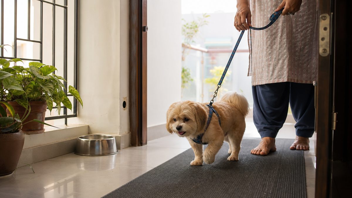
- Lay rubber-backed mats or runners along the routes the dog uses, because tile and polished stone make a dog with a new false joint splay and catch itself.
- Split the walk into short frequent sessions before sunrise and after dark rather than one long one, which is the only way to get controlled exercise into a humid Indian day.
- Book the physiotherapy before the surgery, since rehabilitation begins within the first two weeks and finding a provider afterwards wastes the window.
- Weigh the dog every fortnight on the same bathroom scale, because a smaller dog asks less of a false joint and weight is the one variable you control daily.
What the dog is like at the end of it is worth stating plainly. Most dogs regain very good use of the affected leg and return to normal activities, the removed femoral head does not grow back so the source of the pain is gone for good, and a minority keep a slight intermittent lameness. In the Giessen series, occasional problems showed up after heavy activity, after a long rest, or at a change in the weather, and no owner thought their animal's quality of life was restricted by them.
Unlike hip dysplasia in dogs, which is managed for years, Legg-Calve-Perthes disease has an end point: the diseased bone is removed and the disease does not come back in that hip. The prognosis following appropriate treatment is very good and most dogs go on to comfortable, active lives. What you are buying with early diagnosis is less muscle wasting to rebuild and a shorter recovery. Your nearby vet decides the plan for your dog.
Keep a one-page record from the day the limp starts: the date, the weight every fortnight, the drug classes prescribed, the surgery date, and the week the dog first put the foot down unprompted. That sheet is what lets your vet tell a bad week from a real setback, and only you can collect it. If you have another small-breed dog at home, our guide to adult dog care in India covers the weight and exercise routine that keeps the good leg good.
Questions Google India asks about Legg-Calve-Perthes disease in dogs
How is Legg-Calve-Perthes disease treated in dogs?
Treatment is surgical in most dogs. The femoral head and neck are removed in an operation called an FHO, and the body builds a fibrous false joint from the surrounding muscle, which ends the painful bone-on-bone contact. Rest and anti-inflammatory pain relief are used first as a four-week trial, and surgery is indicated if that trial produces no clinical improvement.
How is Legg-Calve-Perthes disease treated?
The same way in a dog as the question above, by removing the dead femoral head and neck so a false joint can form, with a total hip replacement as the alternative in larger dogs. In children the disease is managed very differently, with bracing and osteotomies that preserve the femoral head, which is why a human treatment page does not transfer to your puppy.
How much does Legg-Calve-Perthes Disease surgery cost for dogs?
In India a femoral head ostectomy is priced at Rs 25,000 to Rs 60,000 by a private clinic chain. A government veterinary college charges by category instead: Madras Veterinary College lists a major small-animal surgery at Rs 3,000 and Bihar Animal Sciences University at Rs 2,500. Add the diagnostic radiograph, Rs 150 at ICAR-IVRI to Rs 5,000 privately, and ask whether sedation is separate.
What are the first signs of Perthes disease?
A limp on one hind leg in a small-breed dog under a year old that gets worse over weeks rather than better. In 35 dogs studied clinically, muscle atrophy was recorded in 25, pain on passive movement of the hip in 28, a shortened leg in 14 and crepitation in 8. Owners usually notice the dog refusing to jump onto furniture first.
Can Legg-Calve-Perthes disease heal on its own?
No. The dead bone does not come back to life, and there is no cure that restores a collapsed femoral head, which is why the operation removes it instead. One review reports that only about 25 per cent of patients respond to conservative treatment, and those that do carry an increased long-term risk of arthritis and some functional impairment.
Is Legg Calve Perthes disease hereditary in dogs?
Partly, and nobody can yet name the gene. A test mating of two affected Manchester Terriers produced three affected males and two unaffected females, and pedigree analysis indicated high heritability. Cornell researchers have narrowed the search to a region on canine chromosome 6 using 85 affected and 102 healthy dogs. The OFA states the mode of inheritance is not known, and every authority advises against breeding affected dogs.
Can both hips be affected?
Yes, in a minority. The only laterality proportion published for dogs puts bilateral disease at 12 to 16.5 per cent of affected animals, citing the two classic canine series. Cornell, PDSA and VCA all describe the disease as usually affecting one hip, while the MSD Veterinary Manual states the lesion is often bilateral, so ask for both hips to be radiographed.
How long does recovery take after FHO surgery in a dog?
About three months of active rehabilitation. In 27 dogs randomised after femoral head and neck excision, the swimming group reached functional recovery at a median of three months and the non-swimming group at six. Physiotherapy starts within the first two weeks, aiming to restore hip extension, and most dogs are walking and trotting normally by three to four months.