
Dog health: ophthalmic
Glaucoma in dogs: the hours that decide whether the eye sees again
A cited, vet-reviewed page on the eye disease that takes sight in under a day. It carries the salvage window nobody prints, breed prevalence with denominators, what gonioscopy actually grades, the three names one screening scheme goes by, and where in India a dog's eye pressure can be measured.
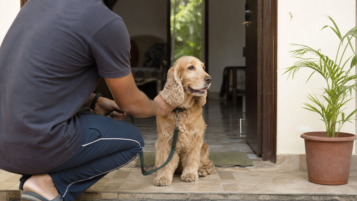
Glaucoma is an emergency. Go to a veterinary clinic now, and call ahead, if you see any of these signs.
- A painful, cloudy, red eye that was normal yesterday
- A widely dilated pupil that does not react to light
- Squinting or holding one eye shut, with obvious pain
- Sudden loss of vision on one side
- An eye that looks visibly bigger than it was
- A quiet, withdrawn dog refusing food alongside any of these
Quick answer
Glaucoma in dogs is raised pressure inside the eye that kills the optic nerve, and the acute form is a same-day emergency. Vision may still be salvageable if the dog is treated while the pressure has been up for less than 12 to 24 hours, so a cloudy, red, painful eye goes to a vet tonight. Risk is concentrated in a few breeds, the American Cocker Spaniel highest at 5.52 percent. No treatment reverses the damage, and the second eye usually follows.
This guide is general information for Indian pet owners and is not a diagnosis. Always consult a registered veterinarian near you before acting on anything you read here, and treat any sudden change in your pet's condition as urgent.
Signs of Glaucoma
The 8 signs to watch for are listed below. Any one of them is a reason to consult your nearby vet; none of them is a diagnosis.
- A cloudy or bluish haze over the surface of one eye
- Redness in the white of the eye
- Squinting, excessive blinking, or rubbing the eye on furniture
- A pupil that stays wide and does not shrink in light
- Pupils of two obviously different sizes
- A dog gone quiet, withdrawn or off its food
- One eye that looks larger than the other
- Bumping into things, or missing a thrown ball on one side
Breeds on this site that document this condition
Each breed page below carries the figure for this condition with its denominator and its source. The 10 breeds listed are the ones on this site that document it, not a ranking of risk.
Tonometry is the Indian constraint, because glaucoma cannot be diagnosed without a tonometer and a tonometer is specialist equipment rather than standard kit in a general practice. Four Indian institutions appear in the published record with one: Kerala Veterinary and Animal Sciences University measured twelve glaucomatous dogs at Mannuthy and Kokkalai with an applanation tonometer, and DUVASU Mathura with RAJUVAS Bikaner measured eighteen working dogs of the Sashastra Seema Bal at Alwar with both a rebound and an indentation tonometer, finding a small but significant 0.76 mm Hg difference between the two instruments. Madras Veterinary College under TANUVAS lists ophthalmology among its surgical referral units, runs a dedicated ophthalmology theatre performing iridectomy among other eye procedures, and publishes rupee charges, although three of its own pages print three different surgery fee tables. India has no prevalence study of canine glaucoma at all, no national eye screening scheme, and no laboratory selling a breed-specific glaucoma DNA test, so an Indian owner reads North American and Japanese denominators and posts a cheek swab abroad in dollars.
What glaucoma in dogs is, and how common it actually is
Glaucoma in dogs is raised pressure inside the eye that destroys the optic nerve and the retina, and in the dog it is a leading cause of irreversible vision loss. The eye makes fluid continuously and drains it through a sieve at the base of the iris; when that drainage angle stops working the pressure climbs, and the nerve dies under it.
There are two questions that matter before any other, and the ranking pages blur them. Is this primary or secondary, and is it open angle or closed angle? Primary glaucoma is an inherited fault in the drainage angle itself and it is the commonest type in dogs; secondary glaucoma is pressure raised by something else in the eye, such as a cataract, a tumour or inflammation. Within primary glaucoma, the angle is either physically closed, which the Royal Kennel Club calls primary closed angle glaucoma and says is the more common of the two in dogs, or it is open with the resistance sitting deeper, which is primary open angle glaucoma.
That split decides everything else on this page, because the two types behave nothing alike. Closed angle disease arrives as a sudden, painful, sight-threatening attack, and the pressure reaches the 50 to 80 mm Hg range in a process that can occur in the span of minutes. Open angle disease is the opposite. The Kennel Club describes it as normally seen in dogs aged between 3 and 6 years, affecting both eyes usually simultaneously, and it states plainly that pain or discomfort is not a normal feature, which is why measurement of the pressure inside the eye is the essential part of any professional examination. One type sends an owner to a clinic at midnight. The other is found only if somebody looks.
Two published prevalence figures exist for North America and they do not agree, which is worth stating because the same author wrote both.
| Source and population | Figure | What it counted | What it does not tell you |
|---|---|---|---|
| Gelatt and MacKay, Veterinary Medical Data Base, North American teaching hospitals, 1994 to 2002 | 0.89 percent | Primary breed-related glaucoma only, in every pure-bred dog presented | Nothing about secondary glaucoma, and nothing about dogs that never reach a teaching hospital |
| The same series, 1964 to 1973 | 0.29 percent | The same diagnosis, thirty years earlier | Whether the rise is real disease or better tonometry |
| MSD Veterinary Manual, written by Kirk N. Gelatt, the author of the series above | about 1.7 percent | Glaucoma in dogs in North America, with no type restriction stated | The denominator, which the page does not print |
| Kato and colleagues, one Japanese referral caseload | 127 of 1,244 dogs | Every dog given tonometry and gonioscopy, 29 breeds | A population rate, because a referral caseload over-represents sick eyes |
The gap between 0.89 percent and 1.7 percent is not sloppiness, it is scope. The lower figure counts primary breed-related glaucoma in pure-bred dogs and it rose steadily across the four decades of the series, from 0.29 percent in 1964 to 1973, to 0.46 percent, to 0.76 percent, and to 0.89 percent by 1994 to 2002. The higher figure is the same author writing for owners about glaucoma in dogs generally, which includes the secondary cases. Read either one as a hospital caseload rate rather than a population rate, because both count dogs that were taken to a veterinary school.
The same breeds, two registers, and a 55-fold disagreement
A second register exists and it disagrees with Gelatt by up to two orders of magnitude, which is worth understanding before trusting either. The register rounds its percentages to one decimal, so several breeds print as 0.0 percent on counts that are not zero, and the recomputed values below are ours rather than the register's. The Orthopedic Foundation for Animals runs an eye-examination register in which a board-certified ophthalmologist records findings on dogs presented for breeding certification, and its glaucoma register holds 29 breed rows. Where Gelatt's teaching-hospital series puts the American Cocker Spaniel at 5.52 percent, that register records 12 affected dogs out of 17,029 Cocker Spaniels examined, or 0.1 percent Orthopedic Foundation for Animals. Neither number is wrong.
| Breed | Gelatt, teaching-hospital caseload 1994 to 2002 | OFA eye register, dogs examined for breeding certification | Ratio of the two, recomputed here |
|---|---|---|---|
| Cocker Spaniel (American) | 5.52 percent | 12 of 17,029, printed as 0.1 percent, 0.07 recomputed | About 78 times higher in the hospital series |
| Shar Pei | 4.40 percent | 2 of 309, printed as 0.6 percent, 0.65 recomputed | About 7 times |
| Siberian Husky | 1.88 percent | 4 of 15,363, printed as 0.0 percent, 0.03 recomputed | About 72 times |
| Cairn Terrier | 1.82 percent | 1 of 2,149, printed as 0.0 percent, 0.05 recomputed | About 39 times |
| Boston Terrier | 2.88 percent | 1 of 10,498, printed as 0.0 percent, 0.01 recomputed | About 300 times |
| Shiba Inu | Not among the ten highest | 2 of 3,243, printed as 0.1 percent, 0.06 recomputed | No hospital-series figure to compare; the Japanese caseload is the better comparison |
Two things explain the whole gap and neither is an error. The eye register examines young dogs and glaucoma is a middle-aged disease. Dogs go through breeding certification as adults of breeding age, while the glaucomas present for first diagnosis between 4 and 10 years in most breeds, so a certification examination is usually taken before the disease would have appeared. And a teaching hospital sees sick eyes while a breeding register sees selected ones. Read the eye register as the chance that a breeding dog already has visible glaucoma on the day it is certified, and read Gelatt as the chance that a dog of that breed reaching a veterinary school is there for glaucoma. An owner asking what their own dog's lifetime risk is has neither number, because nobody has measured that in any country.
The register does still rank breeds, and the ranking is informative. The highest rates in it are the Kai Ken at 1 of 73, the Basset Fauve de Bretagne at 2 of 358 and the Chinese Shar-Pei at 2 of 309, then the Petit Basset Griffon Vendeen at 2 of 757. Three of those four are the ADAMTS17 breeds, which is the genetics showing through even in a population screened too young to have much disease. At the other end, the Labrador Retriever sits at 4 affected in 101,680 examined and the Golden Retriever at 3 in 126,200, and those two denominators are the largest on the whole register.
Why the breed matters more than the average
The average is close to useless for an individual dog, because the risk is concentrated in a small number of breeds. In the last observation period of Gelatt's series, 22 different breeds had a 1 percent or higher prevalence of the glaucomas, and the American Cocker Spaniel topped the list at 5.52 percent. MSD puts the same point more bluntly, that the frequency of breed-predisposed glaucoma in both eyes in purebred dogs is the highest of any animal species except humans. The breed cards on this page carry those numbers one by one.
The Japanese picture is a better guide for an Indian owner than the North American one, because the breed mix is closer. Of 1,244 dogs of 29 breeds given a full examination including tonometry and gonioscopy, glaucoma was diagnosed in 127 dogs and 162 eyes, of which 129 eyes were primary and 33 secondary. The Shiba Inu accounted for 42 of the 127 dogs, 33 percent, the highest of any breed, and the Shih Tzu came second with 21 dogs, 16.5 percent. Every affected Shiba Inu had primary glaucoma with an abnormal iridocorneal angle and a dysplastic pectinate ligament. Shih Tzus are kept in large numbers in Indian cities, and no page written for an Indian reader has put that number in front of them.
Age gives the last piece of the shape. Across the breeds in the North American series, the glaucomas were presented for initial diagnosis in dogs between 4 and 10 years of age in the majority of breeds. In the multicentre study of dogs with closed angle disease in one eye, the mean age at first diagnosis was 8.3 years and 69.2 percent of the dogs were female. This is a middle-aged and older dog's disease, which is why it belongs alongside our guide to senior dog care in India.
The hours that decide whether that eye ever sees again
Acute glaucoma is defined by the clock, not by how bad the eye looks. A specialist reference defines acute glaucoma as an elevation in intraocular pressure of less than 12 to 24 hours' duration, and states that if patients are treated during this phase, vision may be salvageable. Past that window the question stops being whether the dog sees and becomes whether the eye can be made comfortable.
Nothing else on this page is as important as that sentence, and no page written for owners prints it. VCA tells an owner that acute glaucoma is an emergency. Cornell says immediate treatment is needed to save the dog's vision and that eye pressures above 40 to 50 mm require emergency treatment. The Royal Kennel Club says that if the intraocular pressure is not reduced quickly, the eye can be damaged irreversibly. All three are correct and none of them tells the owner how long quickly is. It is under a day.
The speed comes from the mechanics. When the angle closes, aqueous fluid is trapped and pressure builds until it is counterbalanced by the resistance of the uvea, cornea and sclera, usually in the 50 to 80 mm Hg range, and this entire process can occur in the span of minutes. A normal dog's eye sits far below that. Cornell gives normal canine eye pressures as typically a maximum of 20 to 28 mm Hg, and eighteen healthy Indian working dogs measured with a rebound tonometer ranged from 17 to 22 mm Hg with a mean of 19.39 mm Hg. A dog in an attack is carrying two to four times the pressure its optic nerve was built for.
Severe pain is a common feature of most phases of closed angle glaucoma in dogs where the pressure rise is marked, although very mild rises of 5 to 15 mm Hg may cause no ocular pain at all. Owners rarely read it as eye pain. They report a dog that has gone quiet, that will not eat, or that seems depressed, which MSD lists among the behavioural changes of the disease.
Do not wait until morning to see whether a cloudy, red, painful eye settles. The salvage window for an acute attack is under 12 to 24 hours from the pressure rising, and treatment inside it is what makes vision salvageable. Nothing you can buy over a counter lowers eye pressure, and a human eye drop bought at a chemist is not a substitute for a pressure measurement. Go to a veterinary clinic tonight, ask specifically for the pressure in both eyes to be measured, and ask to be referred onward if the practice has no tonometer. Your own vet decides what happens next for your dog; this page cannot.
What an owner can actually see at home
The visible signs are the front of the eye, because the damage at the back is invisible without instruments. The Royal Kennel Club's own list of what an owner may notice runs to nine items, and it puts the behavioural change first rather than last. Six of them are worth memorising if you own one of the breeds on this page.
- A cloudy or bluish haze across the surface of the eye, which is the cornea swelling under pressure.
- Redness in the white of the eye, from congested vessels on its surface.
- Squinting, excessive blinking, or rubbing the eye on furniture.
- A pupil that stays wide and does not shrink in light, or two pupils of different sizes.
- A dog that has gone quiet, withdrawn or off its food, which is how eye pain usually presents.
- An eye that looks larger than the other one, which means the pressure has already been high for some time.
One sign is a warning rather than an emergency, and telling them apart saves an eye. A midrange to dilated, sluggish to fixed, irregularly shaped pupil is a common sign of primary glaucoma in dogs, and owners can sometimes recall episodes of intermittent angle closure on careful questioning. A dog that had a cloudy, sore eye for an evening three weeks ago and then seemed fine has probably already had an attack that resolved on its own. That dog needs its pressure measured this week, not the next time it happens.
An eye that has been under pressure for weeks looks different again, and by then the news is worse. A chronically glaucomatous eye becomes enlarged, which vets call buphthalmia, and such an eye is irreversibly blind. Linear breaks in the innermost corneal layer, called Haab striae, appear as the globe stretches. An enlarged eye is not an early sign. It is a late one, and it changes the goal of treatment from saving sight to ending pain.
How glaucoma in dogs is diagnosed, and what each instrument finds
To diagnose glaucoma a veterinarian measures the pressure inside the eye with a tonometer, and no examination without that measurement can rule the disease in or out. MSD puts it in one line, that a vet uses a special tool called a tonometer to measure the pressure inside the eye. Everything else in the work-up tells you which kind of glaucoma it is and what to do about it.
Three instruments do three different jobs and they are constantly confused. Tonometry gives a number in millimetres of mercury and answers the question of whether there is glaucoma today. Gonioscopy puts a lens on the cornea and looks sideways into the drainage angle to grade how much of it is malformed, which answers whether a dog with a normal pressure is at risk of glaucoma later. Ophthalmoscopy looks at the back of the eye and grades the damage already done to the optic nerve.
Tonometry itself comes in three forms and they do not give the same answer, which matters when a dog is rechecked at a different practice. Indentation, applanation and rebound tonometers are all non-invasive. Each device has an inherent 1 to 2 mm Hg variability according to its manufacturer, but in manometric studies all tonometers can occasionally produce estimates that vary by 3 to 4 mm Hg when applied at exactly the same place at exactly the same true pressure. A clinician should therefore not over-interpret small differences between readings.
The Indian working-dog study makes the same point with its own numbers, and it is the only Indian measurement of this that exists. Eighteen healthy dogs were measured with both a rebound tonometer and an indentation tonometer at the same visit; the rebound instrument read 17 to 22 mm Hg with a mean of 19.39 mm Hg and the indentation instrument read 14.60 to 20.60 mm Hg with a mean of 18.63 mm Hg. The gap of 0.76 mm Hg was small but statistically significant, with the rebound tonometer reading higher. Ask which instrument was used and keep to the same one for rechecks.
What the numbers mean, from three independent sources
Three thresholds get quoted and it helps to see them together rather than one at a time.
| Figure | Value | Population and instrument | Source |
|---|---|---|---|
| Normal canine range | Typically a maximum of 20 to 28 mm Hg | Stated for dogs generally, instrument not specified | Cornell Riney Canine Health Center |
| Normal in healthy Indian working dogs | 17 to 22 mm Hg, mean 19.39 | 18 Belgian Shepherds, German Shepherds and Labrador Retrievers aged 3.5 to 6 years, rebound tonometer | Nema and colleagues, DUVASU Mathura and RAJUVAS Bikaner |
| The same dogs, other instrument | 14.60 to 20.60 mm Hg, mean 18.63 | The same 18 dogs, indentation tonometer at the same visit | Nema and colleagues |
| Emergency threshold | Above 40 to 50 mm | Stated as requiring emergency treatment | Cornell Riney Canine Health Center |
| Typical pressure in an angle-closure attack | 50 to 80 mm Hg | The mechanical ceiling set by the eye's own resistance | Miller and Bentley |
| Pressure in glaucomatous Indian dogs at presentation | Mean 33.99 mm Hg, falling to 18.05 by day 42 on treatment | 12 dogs, applanation tonometer, Kerala university hospitals | Raut and colleagues, KVASU Mannuthy |
One number in that table does a job none of the others can. The Kerala series measured both eyes of every dog, and the unaffected fellow eye read a mean of 18.54 mm Hg on the day the affected eye read 33.99. The dog's own other eye is the control, which is why a vet should always measure both even when only one looks wrong.
The back of the eye is examined next, and in a proportion of dogs it cannot be. In the Kerala series, direct and indirect ophthalmoscopy could not be performed in 5 of the 12 dogs because the cornea or the lens was not clear enough to see through, owing to pigmentary keratitis or cataract. Of the dogs that could be examined, optic disc cupping was seen in two, retinal vessel attenuation in one, cataractous change in four, retinal haemorrhage in one and no change at all in one. A clear view is not guaranteed, and a vet who cannot see the back of the eye is not being careless.
Vision tests are cruder than owners expect and they are read alongside the pressure rather than instead of it. In the same Kerala dogs the menace response was present in 38 percent of affected eyes and absent in 62 percent, while the pupillary light reflex was present in 69 percent. A dog that still blinks at a moving hand can have a pressure of 34 mm Hg. Do not use your own vision test at home to decide whether to go.
Gonioscopy, the screening test nobody explains
Gonioscopy is the only test that finds the risk of primary closed angle glaucoma before the pressure ever rises. An eye panellist puts local anaesthetic drops on the eye and rests a goniolens on the cornea so the drainage angle can be looked at directly in both eyes, and some dogs need sedation for it. What the panellist grades is how much of that angle is blocked by malformed tissue called the pectinate ligament.
The result is a grade rather than a pass or a fail, and it is the only part of the British scheme that works that way. Every other condition on the certificate is recorded as Clinically Unaffected or Clinically Affected, except for the results of gonioscopy, which are graded. The scheme is open to pedigree and crossbreed dogs alike and does not require kennel club registration, and a routine eye examination for one dog starts from GBP 72.00 including VAT, with litter screening for puppies aged 5 to 12 weeks starting from the same figure.
| Grade | How much of the drainage area is abnormal | What the scheme says it means |
|---|---|---|
| Grade 1 | 25 percent or less | Considered normal or mildly affected |
| Grade 2 | Between 26 and 75 percent | Considered moderately affected |
| Grade 3 | More than 75 percent | Considered severely affected and at risk of developing glaucoma |
A second grading scale exists in the research literature and it does not line up with that one. The studies that measured pectinate ligament dysplasia in British breeds classified eyes as unaffected at 0 percent, mildly affected below 20 percent, moderately affected at 20 to 90 percent and severely affected above 90 percent Oliver JA, Ekiri. A dog graded moderately affected under the scheme's 26 to 75 percent band and a dog called moderately affected in a paper's 20 to 90 percent band are not the same dog. Read the percentage, not the word.
Two examiners, the same 98 eyes, and how far apart they were
The limitation nobody tells breeders about is that gonioscopy is an estimate made by eye. Two examiners performed gonioscopy in 98 eyes of 49 Welsh Springer Spaniels and each estimated the percentage of the angle affected to the nearest 5 percent. Their results correlated strongly at R = 0.91, but Examiner 1 generally scored eyes higher than Examiner 2, especially in eyes where both agreed there was an abnormality. A good level of agreement could only be reached with a scheme of no more than three categories and a relatively large intermediate band, at a kappa of 0.68. The authors' own conclusion is that such a scheme would only reliably add the detection of dogs with marked progression, the ones most at risk of closed angle glaucoma.
The second limitation is that the grade moves. Gonioscopy was repeated over time in 65 Welsh Springer Spaniels and 35 of them, 53.8 percent, showed progression of pectinate ligament dysplasia. The authors state the consequence directly, that dogs deemed unaffected at an early age may subsequently be diagnosed with the condition. A clear gonioscopy at eighteen months is a statement about that dog at eighteen months.
The prevalence figures themselves are the third surprise, because screening has not fixed the problem. In a single prospective study, 139 of 227 Welsh Springer Spaniels, 61.2 percent, were affected at grades 1 to 3, and 82 of 227, 36.2 percent, were moderately or severely affected. Gonioscopy in three other British breeds found 76 of 198 Basset Hounds (38.4 percent), 36 of 170 Flatcoated Retrievers (21.2 percent) and 21 of 95 Dandie Dinmont Terriers (22.1 percent) moderately or severely affected. The prevalence was significantly higher in the Basset Hound than in either of the other two, and in all three breeds it rose with age. The authors note the prevalence remained high despite widespread screening and selection against the condition.
The scheme has three names and two different breed lists
Anyone trying to follow the British scheme from India hits a naming problem that took three documents to untangle. The British Veterinary Association's own Eye Scheme page, opened on 24 September 2026, presents it as the Eye Scheme within the Canine Health Schemes, names the Royal Kennel Club as the body that publishes the results, and contains no mention of the ISDS at all. The Royal Kennel Club's own glaucoma article calls the same thing the BVA/KC/ISDS Eye Scheme. The Royal Kennel Club Health Standard of February 2026 calls it the BVA/RKC/ISDS Eye Scheme, 143 times across 33 pages. Three names, two organisations, one scheme. If you are asking a breeder for a certificate, ask for a gonioscopy grade and a date rather than for a scheme by name.
The breed lists disagree too, and the disagreement has consequences. The Health Standard asks for eye testing and gonioscopy for pectinate ligament abnormality in 14 breeds, including the Basset Hound, Border Collie, Dandie Dinmont Terrier, Hungarian Vizsla, Japanese Shiba Inu, Leonberger, Flat Coated Retriever, Golden Retriever, Siberian Husky, American Cocker Spaniel, Cocker Spaniel, English Springer Spaniel, Welsh Springer Spaniel and Spanish Water Dog. Set that against Gelatt's ten highest-prevalence breeds and only three of the ten appear on it, namely the Basset Hound, the Siberian Husky and the American Cocker Spaniel The Royal Kennel Club Health Standard. The Chow Chow at 4.70 percent, the Boston Terrier at 2.88 percent, the Wire Fox Terrier at 2.28 percent, the Cairn Terrier at 1.82 percent and the Miniature Poodle at 1.68 percent are each asked for eye testing under the scheme but for no gonioscopy. That is a British standard built around British breed clubs, and it is not a ranked list of risk.
The DNA tests, which genes they cover and which breeds they do not
A DNA test for glaucoma exists for one type only. The Royal Kennel Club states that both primary closed angle and primary open angle glaucoma have a hereditary component in some breeds, but that only primary open angle glaucoma, in some breeds, has a DNA test available. Closed angle disease, the more common type in dogs, has no test and is screened by gonioscopy instead.
Two genes carry almost all of the tested disease and they split the breeds cleanly between them. Variants in ADAMTS10 are documented in the Beagle and the Norwegian Elkhound, and variants in ADAMTS17 in the Basset Hound, the Basset Fauve de Bretagne, the Chinese Shar-Pei and the Petit Basset Griffon Vendeen. Each breed has its own causal variant rather than a shared one, which is why a test validated for one breed says nothing about another.
The variant hunt in each breed produced a different kind of mutation, and the detail is the reason the tests are breed-specific. In the Basset Hound it is a 19 base-pair deletion in exon 2 that truncates the protein; in the Basset Fauve de Bretagne a missense change in exon 11; in the Petit Basset Griffon Vendeen a 4.96 megabase inversion with one breakpoint inside the gene; and in the Chinese Shar-Pei a 6 base-pair deletion in exon 22 Forman OP, Pettitt L, Komaromy AM. In the Norwegian Elkhound it is a single missense change in exon 9 of ADAMTS10. All of them are inherited as autosomal recessive traits, so a dog needs two copies to be affected and one copy makes it a carrier that stays healthy but passes the variant on.
Two of those studies published a carrier frequency in the wider breed, and both numbers are high enough to change breeding decisions. Genotyping 223 Basset Hounds recruited for an unrelated study, and therefore a random sample of the United Kingdom population, gave a mutant allele frequency of 0.081 and a predicted frequency of affected dogs of 0.007. In the Norwegian Elkhound the missense variant was found in 14 cases against 572 unaffected dogs with a carrier frequency of 25.3 percent, meaning roughly one Elkhound in four carries it.
What the world's DNA register actually holds
The register that records these results is small, and its size is the most telling number on this page. A live reading of the Orthopedic Foundation for Animals disease statistics on 24 September 2026 shows that its primary open angle glaucoma register holds 632 evaluations in total across all breeds, while its primary lens luxation register holds 17,677. The test for the blinding glaucoma has been run roughly twenty-eight times less often than the test for a related lens disease in the same terriers. Every row below was arithmetic-checked against its own three percentages and each sums to exactly 100.0.
| Register and breed | Evaluations | Normal | Carrier | Abnormal |
|---|---|---|---|---|
| Primary open angle glaucoma, Petit Basset Griffon Vendeen | 316 | 53.8 percent | 43.4 percent | 2.8 percent |
| Primary open angle glaucoma, Beagle | 139 | 99.3 percent | 0.7 percent | 0.0 percent |
| Primary open angle glaucoma, Basset Hound | 83 | 100.0 percent | 0.0 percent | 0.0 percent |
| Primary open angle glaucoma, all breeds combined | 632 | 74.7 percent | 23.7 percent | 1.6 percent |
| Primary closed angle glaucoma, Italian Greyhound | 275 | 81.5 percent | 18.5 percent | 0.0 percent |
| Primary closed angle glaucoma, all breeds combined | 303 | 81.8 percent | 18.2 percent | 0.0 percent |
| Shar-Pei open angle glaucoma with lens luxation, Chinese Shar-Pei | 78 | 79.5 percent | 20.5 percent | 0.0 percent |
The Petit Basset Griffon Vendeen row is the one to look at twice. Of 316 dogs tested, 43.4 percent came back as carriers and 2.8 percent as affected, so close to half the tested population of that breed carries the variant. That is consistent with the research history, where the locus was mapped in the breed with only 12 cases and 12 controls because the disease was that easy to find. The Basset Hound row runs the other way, with all 83 tested dogs normal, which is what a breed looks like once a test has been used for a decade.
One finding in this literature deserves to be better known, because it explains how the problem was created. The glaucoma-causing ADAMTS17 variants are also reproducibly associated with height, and affected dogs are significantly shorter than clear dogs of the same breed. In 233 Petit Basset Griffon Vendeens the adjusted mean height at the withers was 33.49 cm in affected dogs against 34.92 cm in clear dogs, and in 66 Shar-Peis it was 43.96 cm against 48.95 cm. The authors' conclusion is that selecting dogs to be of a specific short height may have increased the frequency of these mutations within these breeds. Breeders selected for a look and got a blinding eye disease attached to it.
Where you buy the test matters less than which variant it runs. The Kennel Club sells its primary open angle glaucoma test for the Basset Hound, the Norwegian Elkhound and the Petit Basset Griffon Vendeen, and records a result only from a laboratory on its own approved list. Its own Health Standard names five different POAG test codes across five breeds, adding the Basset Fauve de Bretagne and a combined open angle glaucoma and lens luxation test for the Shar Pei The Royal Kennel Club Health Standard. The University of California Davis Veterinary Genetics Laboratory sells the Petit Basset Griffon Vendeen test at USD 58 for a single animal, from cells brushed from the dog's cheek, with a turnaround of at least 15 business days. There is no Indian laboratory selling a breed-specific glaucoma variant test, so an Indian buyer posts a swab abroad and pays in dollars.
One caution applies to every one of these tests and it is the same one that applies to progressive retinal atrophy in dogs. A clear result clears a dog of one named variant in one gene, and glaucoma in dogs is bilateral, breed-specific and genetically heterogeneous. Evidence that variants in ADAMTS10 are not causal in the Shiba Inu and the Shih Tzu, and that a different gene is involved in those breeds, is recorded in the same registry that documents the Beagle and the Elkhound. A Shiba Inu owner reading an ADAMTS10 result has learned nothing about their own dog.
Treatment for glaucoma in dogs, and the odds each option actually carries
Treatment for glaucoma has one immediate goal and one long one. The immediate goal is to bring the pressure down as fast as possible to try to salvage vision, and the long one is to keep the eye comfortable for as long as it lasts. There is no cure for canine glaucoma and many affected dogs go blind despite a growing spectrum of medical and surgical therapies.
This page names drug classes and never a dose, because the dose is a decision for the veterinarian looking at your dog. Four classes of pressure-lowering eye medication are in routine use and each works differently. Carbonic anhydrase inhibitors reduce the production of fluid inside the eye. Beta-blockers reduce production and also improve outflow, and their pressure-lowering effect on its own is mild, so they are usually combined Reinstein S, DVM MS DACVO. Prostaglandin analogues open the outflow route and can be effective at bringing pressures back down to normal. Hyperosmotic agents given into a vein pull water out of the eye and act within minutes for a matter of hours, and they are used in the emergency itself rather than long term.
Beta-blockers carry a specific caution an owner should know about before a first visit. Undesirable cardiac and respiratory effects can be seen with topical beta-blockers, including a slow heart rate and narrowing of the airways, so they are avoided in dogs with cardiovascular disease and in animals with asthma. Tell the vet about any heart or airway condition before the first drop goes in.
The one Indian treatment series published so far shows what good medical control looks like on real numbers. Twelve glaucomatous dogs seen at two Kerala university veterinary hospitals were treated for 42 days with a combination of a carbonic anhydrase inhibitor and a beta-blocker; mean pressure in the affected eyes fell from 33.99 mm Hg on presentation to 19.76 by day 14, 19.94 by day 28 and 18.05 by day 42, a statistically significant fall, while the unaffected eyes stayed level. The authors' own caveat is the important part, that this treatment is not a cure for glaucoma but a strategy to slow progression and relieve the signs.
Medication also stops working, and owners are rarely warned. In primary glaucoma, medications become less effective over time and the pressure rises again, so combination therapy and increasing frequency are expected rather than a sign of failure. PDSA puts the same thing to owners, that eye drops may stop responding and a new plan then has to be agreed with the vet. The specialist consensus is blunter still, that glaucoma therapies in dogs frequently fail within months with rebounding pressure and blindness.
Surgery, and the published one-year success rates
Surgery divides into operations that try to save sight and operations that end pain in an eye that is already blind. The sight-preserving group is drainage implants that shunt fluid to a reservoir under the conjunctiva, cyclodestructive techniques that reduce fluid production with a laser, and combinations of the two. The salvage group, used in a chronically blind eye, is enucleation, evisceration with an intraocular prosthesis, and chemical ablation of the fluid-producing tissue.
| Procedure | One-year success rate | What success means here | The caveat the authors attach |
|---|---|---|---|
| Ahmed valved drainage implant | About 90 percent | Both pressure control and sight preserved at one year | Improved by better implant design, patient selection and home pressure monitoring |
| Baerveldt non-valved drainage device | 65 to 75 percent | The same combined outcome | Scarring over the drainage bleb is a main reason for implant failure |
| Transscleral cyclophotocoagulation, alone or with an Ahmed implant | 41 to 92 percent | The same combined outcome | The widest range of any option, so the surgeon and the stage matter enormously |
| Endolaser cyclophotocoagulation | 72 to 74 percent | The same combined outcome | Needs intensive postoperative care and considerable expense |
Read that table with the sentence the same authors put under it. Most of the published canine studies are severely limited by small sample size and short follow-up, which is a shortcoming the profession itself has flagged as a priority to fix. Even with the newer methods, they are often associated with intensive postoperative care and considerable expense, and a substantial number of affected dogs still go blind. The choice of method depends on clinician preference and cost to the owner, and on the breed, the type and stage of glaucoma and the surgeon's experience.
Removing the eye is not a failure of care and it is often the kindest option. PDSA states that where glaucoma is not responding to treatment and is causing a lot of pain, a vet may recommend enucleation, that many owners struggle with the idea, and that it is frequently the kindest choice because it prevents further pain, with many dogs coping well afterwards. An eye that is already blind and painful has nothing left to save. A dog with one eye navigates perfectly well.
The second eye, and a disagreement worth knowing about
Primary glaucoma is a bilateral disease, so the healthy-looking eye is the next problem rather than a bystander. How to protect it is genuinely disputed, and both sides are published. A 2018 clinical review states that preventive treatment of the other eye should be recommended and that one beta-blocker delays the onset of glaucoma in it from an average of 8 months to 31 months. A 2026 multicentre retrospective study of 117 dogs with closed angle glaucoma in one eye found that time to failure in the second eye did not differ between dogs given preventive carbonic anhydrase inhibitors and those given beta-blockers, nor between dogs given topical corticosteroids and those not, and concluded that no treatment regimen was superior to another for this purpose.
Those two findings are not actually contradictory, and the distinction is the useful bit. The 2018 figure compares treating against not treating; the 2026 study compares one drug class against another and does not crown a winner among them Donohue LK, Bentley E, Boush JJ. What the 2026 study settles is that arguing about which preventive drop to use is not where the benefit lies. Ask your veterinary ophthalmologist to examine and measure the second eye on a schedule, whatever is prescribed for it.
Natural or over-the-counter treatment is one of the questions Google India asks and it deserves a direct answer. No source consulted for this page describes any dietary supplement, herbal preparation or home remedy that lowers intraocular pressure in a dog, and the specialist consensus paper reviewing the entire therapeutic field lists medical, surgical, gene and stem-cell approaches without any such option. An eye under 50 mm Hg of pressure is a mechanical problem on a clock, and the only things that change it are drugs, a needle or a laser.
Glaucoma in India, where a dog's eye pressure can actually be measured, and what it costs
Tonometry is the constraint in India, because a diagnosis of glaucoma is impossible without a tonometer and a tonometer is a specialist instrument rather than standard equipment in a general practice. The honest answer to where the best glaucoma treatment in India is, is that the documented capability sits in the state veterinary universities, because those are the Indian institutions that have published canine eye-pressure work and that publish their own charges.
Four Indian institutions appear in the published record with a working tonometer, and naming them is safer than naming a clinic list nobody can verify. Kerala Veterinary and Animal Sciences University measured intraocular pressure in twelve glaucomatous dogs at its University Veterinary Hospitals at Mannuthy and Kokkalai using a digital applanation tonometer, over twelve months from December 2021. DUVASU Mathura in Uttar Pradesh, with RAJUVAS Bikaner in Rajasthan, measured eighteen working dogs with both a rebound tonometer and an indentation tonometer. Those are published measurements with instruments named, not claims from a clinic's own marketing.
Geography is the part an Indian owner has to solve for themselves, and it is worth being blunt about the map. The published Indian canine eye-pressure work comes from Thrissur in Kerala, from Mathura in Uttar Pradesh with Bikaner and Alwar in Rajasthan, and the one published eye theatre and fee list comes from Chennai in Tamil Nadu Nema A, Purohit S, Pandey RP, Nema V Tamil Nadu Veterinary and Animal Sciences…. Nothing in that record covers Mumbai, Delhi, Bengaluru, Hyderabad, Pune or Kolkata, which is where most Indian pet dogs actually live. That is an absence in the literature rather than evidence that those cities lack tonometers, and the way to settle it for your own dog is a phone call. Ask the practice one question before you travel, whether it can measure intraocular pressure today and with which instrument. A practice without a tonometer cannot diagnose or rule out glaucoma, however good it is at everything else.
The Chennai picture is the most concrete, because the college publishes both its capability and its prices. Madras Veterinary College in Chennai, part of the Tamil Nadu Veterinary and Animal Sciences University, lists Ophthalmology among the referral units of its Department of Clinics, and its Department of Veterinary Surgery and Radiology runs a dedicated Ophthalmology Theatre Tamil Nadu Veterinary and Animal Sciences…. The eye procedures it names performing there include iridectomy, repositioning of the eyeball, conjunctival flap surgery and keratectomy, alongside cataract extraction by phacoemulsification with lens implantation. Iridectomy is a glaucoma-relevant procedure, and this is the only Indian institution found for this page that names an eye theatre and a fee list on the same site.
The cost in India of the surgical side can therefore be quoted from a government rate card rather than guessed. Every rupee figure below was divided by 84.38 and by 74 before publication and none of them lands on a round dollar amount, so these are Indian prices rather than converted ones.
| Institution and table | Item | Charge | What it does not include |
|---|---|---|---|
| TANUVAS, Madras Veterinary College, Department of Clinics | Small animal registration, valid 7 days | Rs 100 | Any procedure, drug or test |
| The same table | Minor surgery / major surgery / emergency surgery / specialized surgery | Rs 500 / Rs 1,500 / Rs 2,500 / Rs 3,000 | Which category an eye operation falls into |
| TANUVAS, Department of Veterinary Surgery and Radiology, charges approved by its Hospital Management Committee | Minor surgery / major surgery / specialized or emergency surgery | Rs 1,000 / Rs 3,000 / Rs 5,000 | That these differ from the Department of Clinics figures above for the same college |
| TANUVAS, Veterinary University Peripheral Hospital, Madhavaram Milk Colony | Registration for pet animals, valid 1 week | Rs 100 | The peripheral hospital prints its own surgery figures again |
| University of California Davis Veterinary Genetics Laboratory, for comparison | Primary open angle glaucoma DNA test, one dog | USD 58 | Shipping a swab from India, and any Indian equivalent, because none exists |
Two things jump out of that table. The first is that one Indian veterinary college publishes three different surgical fee tables across its own web pages, so a quoted figure needs to be confirmed at the counter rather than read off a website. The second is the comparison at the bottom. A single DNA test posted abroad at USD 58 works out at roughly Rs 4,900 at 84.38 to the dollar, which is about what the same college charges for specialized or emergency surgery. No Indian source found for this page prints a price for tonometry as a line item, and that absence is reported here rather than filled with a guess.
What the Indian research record actually contains
The published Indian evidence on canine glaucoma is thinner than the breed pages might suggest, and it is worth stating exactly how thin. A PubMed search on 24 September 2026 for glaucoma combined with dog, dogs or canine and scoped to India in the title and abstract returned exactly one record, and that record is an artefact: a StatPearls human-ophthalmology chapter on artificial intelligence in the diagnosis of Pythium keratitis, which matches because it mentions dogs, glaucoma and India in different sentences. The same search without the India term returns 669 records, and a control search for parvovirus in Indian dogs returns 40, so the instrument works.
The affiliation search finds different papers and it found the real ones. Searching the same glaucoma terms against Indian author affiliations returned two records, of which one is a genuine Indian canine study and the other is an Australian rabbit tonometry validation with one co-author affiliated in Chandigarh. Widening to intraocular pressure with Indian affiliations returned three, including the Rajasthan working-dog study used throughout this page and, memorably, a human yoga study that matched only because one of the postures it measured is called Downward facing dog. Those are the traps in this literature, and the absence below is published with its controls rather than as a bare claim.
The one Indian record that names glaucoma as a routine finding gives no glaucoma denominator, and that is worth saying plainly. Fifty geriatric dogs were screened at the Teaching Veterinary Clinical Complex of the Orissa University of Agriculture and Technology, Bhubaneswar, between July 2012 and June 2013, with an average age of 10.21 years; glaucoma is listed among the external visible changes commonly encountered in the aged dogs, alongside greying, osteoarthritis and dental wear, but no count of affected dogs is published. The breed spread of those fifty dogs is itself useful for an Indian reader, with the Spitz at 15 dogs, the Indian pariah type at 8, the German Shepherd at 5 and the Labrador at 4.
So the state of Indian evidence is this. India has published one treatment series of 12 dogs and one normal-reference study of 18 dogs, both with named tonometers, and no prevalence study of canine glaucoma at all. An Indian owner is therefore reading North American and Japanese denominators and applying them to an Indian dog, which is defensible for breed risk and not defensible for how common the disease is here. Anyone quoting an Indian glaucoma prevalence figure is quoting something that has not been measured. India also has no national eye screening scheme to put against the British one, so an Indian buyer asking a breeder about glaucoma is asking for private records rather than for a scheme certificate, and a Kennel Club of India pedigree is not a health record; our guide to KCI registration explained sets out what that certificate does and does not cover.
Living with it, the other eye, and the question owners ask first
A dog can live a normal life with glaucoma once the pain is controlled, and the prognosis for comfort is much better than the prognosis for sight. Dogs rank vision third behind smell and hearing, and a dog that is free of pain in a familiar home copes with sight loss far better than its owner expects. The part to plan for is the second eye.
The number every owner of a one-eyed case should know comes from a study of 117 dogs. Within five years, 70.9 percent of dogs with closed angle glaucoma in one eye developed glaucoma in the other eye, and the median time to that happening was 2.15 years. Failure was defined as the second eye passing 25 mm Hg. Dogs whose first diagnosis was in the left eye carried a 1.8-fold higher risk of the right eye failing, which the authors report without explaining. Plan on the second eye, and put the recheck appointments in a calendar rather than waiting for signs.
The breed list in that study reads like an Indian city's dog population. The commonest purebreds among the 117 were the Cocker Spaniel, the Basset Hound and the Shih Tzu. Cocker Spaniels and Shih Tzus are kept in large numbers across Indian cities, which makes this the most directly transferable finding on the page.
Most dogs with primary glaucoma do eventually lose sight in both eyes, and PDSA says so to owners without dressing it up. Glaucoma cannot be cured, although most cases can be well managed with medication for a period. The Royal Kennel Club draws the same line, that unless the glaucoma developed because of another condition, it cannot be cured and needs long-term management. Living with the disease means managing it, not beating it.
Where the dog is going blind slowly, the practical adjustments are the same ones that work for any losing-sight dog, and several of them are free.
- Leave the furniture where it is, because a dog with failing sight navigates from memory.
- Keep food and water bowls in one fixed place, every day.
- Gate the stairs and fence any open water tank or terrace edge, which matters in Indian flats and terraces.
- Teach voice commands now, while the dog can still see you give them.
- Use a harness and lead in any unfamiliar place, including on travel.
- Keep the recheck appointments for the second eye, because it fails at a median of 2.15 years.
When to put a dog down with glaucoma
Blindness on its own is not a reason to put a dog down, and no source consulted for this page recommends euthanasia for sight loss from glaucoma. The relevant question is pain, not vision, and there is a surgical answer to pain before there is a final one. Where an eye is not responding to treatment and is causing a lot of pain, removing it is often the kindest option precisely because it prevents further pain, and many dogs cope really well afterwards. That decision comes before any conversation about euthanasia, not after it.
A dog that is blind, comfortable, eating and interested in its walk has a good quality of life, and the disease itself does not shorten it. The things that change the answer are uncontrolled pain in an eye that cannot be saved or removed, and a dog that has stopped eating, sleeping or engaging. Contact your vet and ask two specific questions, what the pressure in each eye is today, and whether removing the painful eye would make this dog comfortable. Only the veterinarian examining your dog can judge its quality of life, and our guide to when your pet dies in India covers what follows if the answer is the hard one.
One last piece of practical planning is worth flagging early rather than late. Lifelong medication, repeat specialist visits and the real possibility of surgery in both eyes make this an expensive disease to carry, and the specialist consensus explicitly calls for more affordable treatment options as an unmet need. If you own one of the breeds carded on this page, read our guide to pet insurance in India before a diagnosis rather than after one, and ask a breeder for a dated gonioscopy grade using the checks in dog breeder checks in India.
What Google India asks about glaucoma in dogs
Can a dog live a normal life with glaucoma?
Yes, once the pain is controlled. Dogs rank vision behind smell and hearing, and a comfortable dog in a familiar home adapts well to losing sight. The disease itself does not shorten life. What needs planning is the second eye, which failed within five years in 70.9 percent of 117 dogs studied.
What are the first signs of glaucoma in dogs?
A cloudy or bluish haze on the eye surface, redness in the white, squinting or rubbing, a pupil that stays wide in light, and pupils of different sizes. Behaviour often changes first, with a dog going quiet, withdrawn or off its food because the eye hurts. An enlarged eye is a late sign.
Can glaucoma in dogs be cured?
No. Glaucoma in dogs cannot be cured and it requires long-term management, unless it developed because of another treatable eye condition. Specialists reviewing the whole field state there is no cure and that many affected dogs go blind. Medication controls pressure for a time and often becomes less effective as years pass.
When to put a dog down with glaucoma?
Blindness alone is not a reason. The question is uncontrolled pain, and there is a surgical answer before a final one. Where an eye is not responding and hurts, removing it is often the kindest option because it prevents further pain, and many dogs cope well afterwards. Your own vet judges quality of life.
What is the most effective treatment for glaucoma in dogs?
Nothing works for every dog. Among surgical options, published one-year rates for both pressure control and keeping sight are about 90 percent for Ahmed valved drainage implants, 65 to 75 percent for Baerveldt devices, 41 to 92 percent for transscleral cyclophotocoagulation and 72 to 74 percent for endolaser cyclophotocoagulation, all from small studies with short follow-up.
How expensive is glaucoma treatment for dogs?
No Indian source found prints a tonometry price. For scale, Madras Veterinary College publishes small animal registration at Rs 100 and surgery at Rs 500 to Rs 3,000 on one table and Rs 1,000 to Rs 5,000 on another. Lifelong medication and repeat specialist visits are the larger running cost.
Can dogs with glaucoma be treated naturally?
No source consulted for this page describes any supplement, herb or home remedy that lowers a dog's eye pressure. The specialist consensus reviewing the entire therapeutic field covers medical, surgical, gene and stem-cell approaches and lists no natural option. High pressure is a mechanical problem on a clock, and delay costs sight.
Where is the best treatment of glaucoma in India?
The documented capability sits in the state veterinary universities. Madras Veterinary College lists ophthalmology as a surgical referral unit and runs a dedicated eye theatre; Kerala Veterinary and Animal Sciences University and DUVASU Mathura have published canine tonometry with named instruments. Ask any practice directly whether it owns a tonometer.
How is glaucoma in dogs diagnosed?
By measuring intraocular pressure with a tonometer, which no examination can substitute for. Gonioscopy then grades how much of the drainage angle is malformed, and ophthalmoscopy assesses the optic nerve. Instruments disagree by up to 3 to 4 mm Hg at the same true pressure, so keep to one tonometer for rechecks.
Which dog breeds are most affected by glaucoma?
In the largest North American series the highest prevalences were the American Cocker Spaniel at 5.52 percent, Basset Hound 5.44, Chow Chow 4.70 and Shar-Pei 4.40, with 22 breeds above 1 percent. In a Japanese caseload the Shiba Inu and Shih Tzu led instead, which is closer to India's breed mix.